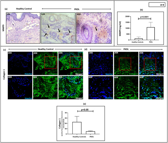
Fig 4. Status of MMP9 and Collagen I in Post Kala-azar Dermal Leishmaniasis (PKDL).
(a): Representative immunohistochemical profiles showing the expression of matrix metalloproteinase 9 (MMP9) in dermal biopsies of a healthy control (n = 6) and patient with PKDL (n = 11) at 10X magnification. Presence of MMP9 has been indicated with black arrowheads. In PKDL, areas with positive staining are further imaged at 40X magnification. (b): Bar graphs indicating the levels of circulatory MMP9 (ng/ml) in healthy controls (n = 10) and patients with PKDL (n = 20). Each horizontal bar represents the median (IQR). (c-e): Representative immunofluorescence profiles showing the expression of collagen I in dermal biopsies of a healthy control (c) and patient with PKDL (d), at 20X and 40X magnification. The red squares in 20X images represent the area that was imaged at 40X magnification. Bar graphs (e) showing the expression of collagen I in healthy controls (n = 7) and patients with PKDL (n = 7). Each horizontal bar represents the median (IQR).

