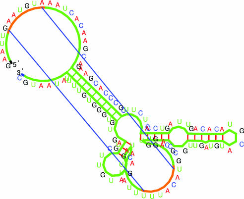
Figure 3.
Illustration of predicted structure including a ‘pseudoknot’ (the helix is visualized by two straight lines corresponding to the first and last base pairs) and an ‘entangled helix’ (base pairs are drawn with the same color as the phosphate-ribose backbone, see main text). The image is generated by the RNAMovies software upgraded to display pseudoknots and entangled helices (38). The structure shown is a co-transcriptional folding intermediate of the 5′-UTR of the infC gene from Bacillus subtilis. The RNA Pol at the nascent 3′ end prevents disentanglement of the entangled helix.

