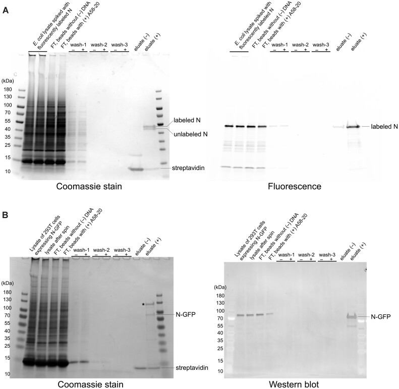
Figure 7.
DNA aptamer-mediated selective pull-down of SARS-CoV-2 N protein. (A) Pull-down from an E. coli lysate spiked with AZDye 488-labeled N protein. The SDS-PAGE gel on the left was stained by Coomassie blue for the total protein, whereas the gel on the right was scanned for fluorescence. Every two lanes show a comparison of results with Streptavidin-coated magnetic beads without (–) and with (+) the biotinylated A58-20 DNA aptamer. ‘FT’ denotes flow through. The Coomassie-stained gel shows that both AZDye 488-labeled and residual unlabeled N protein were pulled down. Based on a fluorescence intensity measurement, 2.1 and 0.16% of the total labeled protein was recovered in (+) and (–) eluate, respectively, in this experiment. (B) Pull-down from the lysate of 293T cells expressing N-GFP. The SDS-PAGE gel on the left was stained by Coomassie blue for the total protein. Immunoblot with an anti-N monoclonal antibody is shown on the right. Representative results of two (A) or three (B) replicates are shown.

