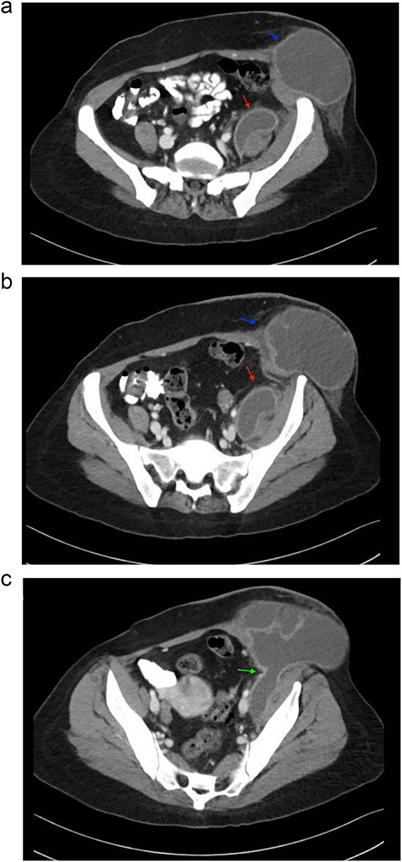
Figure 1.

Sequential images of CT with contrast of the abdomen and pelvis demonstrating the pathology. (a) Slice 96—a small fluid collection in the left psoas muscle (lower arrow), and a low-density fluid collection in the left inguinal region (upper arrow), both with wall enhancement, consistent with abscess formation. (b) Slice 102—progression and enlargement of the iliopsoas (lower arrow) and inguinal (upper arrow) abscesses. (c) Slice 110—connection between the iliopsoas and inguinal abscesses, indicating advanced spread of infection (arrow).
