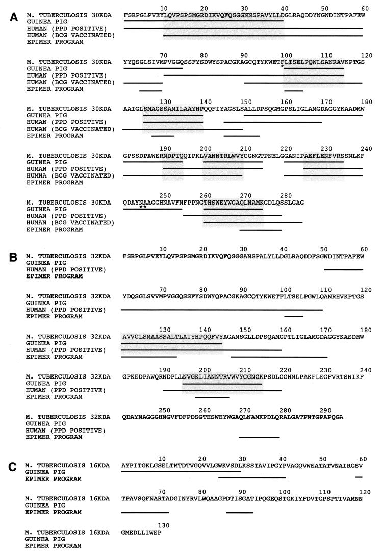
FIG. 2.
Alignment of T-cell epitopes of the 30-kDa (A), 32-kDa (B), and 16-kDa (C) proteins mapped in guinea pigs and in humans. T-cell epitopes that were recognized by at least 25% of the immunized guinea pigs or healthy PPD-positive or BCG-vaccinated individuals are indicated by underlining. Common epitopes of guinea pigs and humans are highlighted with shaded boxes. Epitopes of the 30-, 32-, and 16-kDa protein sequences of M. tuberculosis predicted by the EpiMer computer-based program that are at least five amino acids long are underlined. Epitope mapping in guinea pigs was performed with overlapping peptides derived from the sequence of the 30-, 32-, and 16-kDa proteins of M. tuberculosis shown in the figure. Synthetic peptides used to map the T-cell epitopes of the 30-kDa protein in humans were based on the sequence from M. bovis BCG Tokyo (17, 18). An asterisk indicates a difference in amino acid residue between the 30-kDa protein sequence from M. tuberculosis and that from M. bovis BCG Tokyo (i.e., F100, N245, A246 and L100, K245, P246 for M. tuberculosis and M. bovis BCG Tokyo, respectively). Synthetic peptides used to map the T-cell epitopes of the 32-kDa protein in humans were based on the sequence from M. tuberculosis (9).

