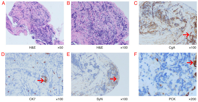
Figure 4.
Histopathological findings of bronchoscopic biopsy and representative IHC-labelled tumor tissue. (A and B) Representative images of tumor morphology. Magnification, ×50 and ×100, respectively. The histological results of (C) CgA, (D) CK7, (E) SyN and (F) PCK. The red arrows indicate positive IHC labelling. IHC, immunohistochemistry; PCK, Pancytokeratin; H&E, hematoxylin and eosin; CgA, Chromogranin A; SyN, Synaptophysin; CK7, Cytokeratin 7.

