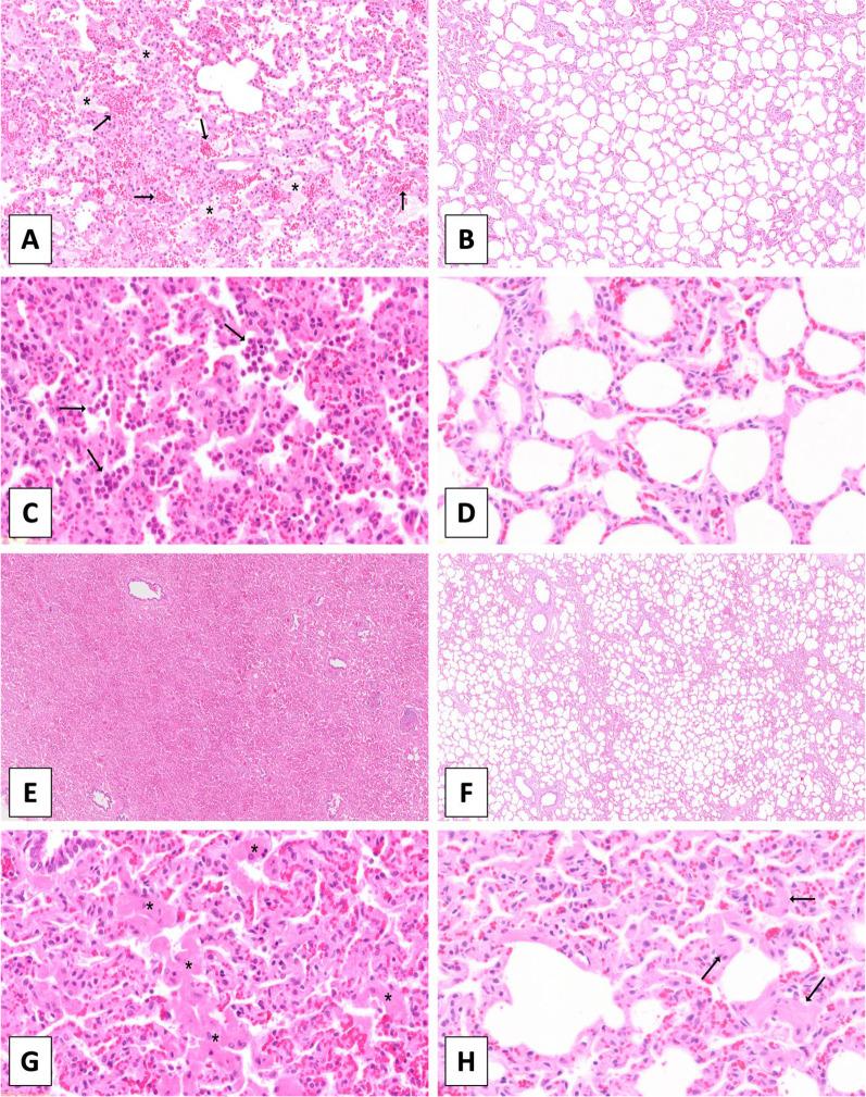
Fig. 4.
Histology images (H&E staining), demonstrating typical histological features. A Presence of mild intra-alveolar edema (asterisks) and hemorrhage (arrows). B Absence of edema or intra-alveolar hemorrhagic material. C Intra-alveolar accumulation of neutrophils (arrows) and mild increase in the numbers of interstitial neutrophils. D Low numbers of neutrophils within the interstitial space. E Severe diffuse atelectasis with alveolar collapse. F Normally aerated alveoli. G Severe multifocal thickening of the alveolar septa caused by hypertrophy of smooth muscle cells (asterisks). (H) Mild septal smooth muscle hypertrophy (asterisks). A, B Magnification 5x. C, D, G, H Magnification 20x. E, F Magnification 2x

