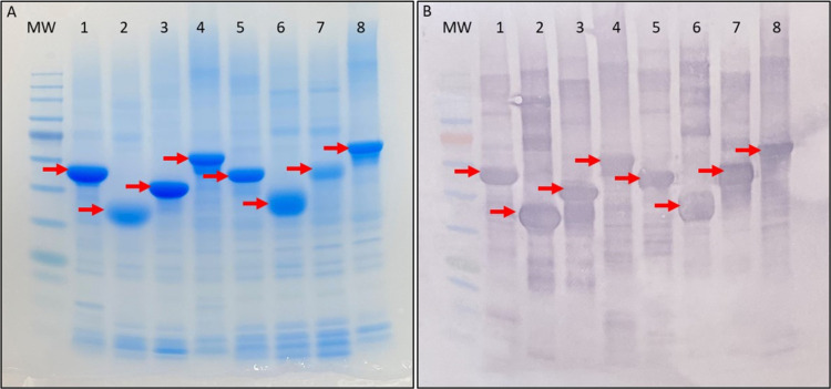
Figure 1.
Analytical protein gel and membrane of the purified constructs (indicated by the red arrows) (a) Coomassie-stained SDS-PAGE and (b) Western Blot using an antihistidine-tag antibody. Lanes are numbered from left to right as follows: MW–protein ladder; 1–N-α; 2–α central rod domain; 3–α-C; 4–N-α-C; 5–N-γ; 6–γ central rod domain; 7–γ-C; 8– N-γ-C.

