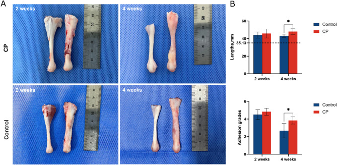
Fig. 2.
(A) Native tendons (left) versus regenerated tendons (right). At 2 weeks, the tenotomy gap connected with insufficient granulated tendon fibrils in the CP group; the tenotomy gap was connected by appropriate granulated tendon fibrils in the control group. At 4 weeks, the tenotomy site of the CP group formed in a dumbbell shape. The tenotomy site of the control group formed in a spindle shape. (B) Length and adhesion grades. The dotted line indicates the mean length of native tendons. Data is represented as mean ± SD. *p < 0.05

