. 2024 Nov 27;12(11):e010069. doi: 10.1136/jitc-2024-010069

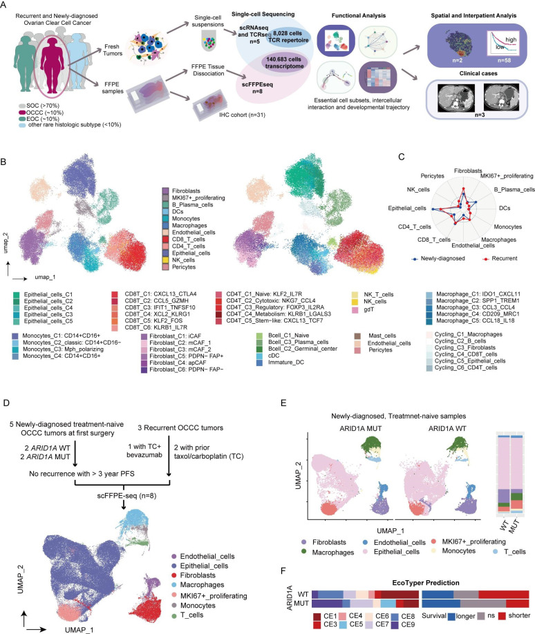
Figure 1. Single-cell multidimensional dissection of OCCC. (A) Study design: five fresh OCCC samples were collected during surgery and underwent single-cell RNA sequencing (scRNA-seq) and single-cell T-cell receptor sequencing (scTCR-seq). Major cell compositions and functional subsets were identified and characterized through integrated analysis. Eight formalin-fixed paraffin-embedded (FFPE) samples with follow-up data were retrospectively collected for scFFPE-seq. Additionally, 31 OCCC samples were used for IHC staining. Analysis on spatial transcriptomics and bulk RNA sequencing datasets were also conducted. (B) Integrative UMAP plot of 49 228 single cell transcriptomics from five harmonized OCCC fresh tumors visualizing main cell compartments (left) and refined cell subsets (right) in OCCC. (C) Radar plot comparing cell proportions in newly diagnosed (blue, n=2) and recurrent OCCC (red, n=3). (D) Flowchart of sample collection for scFFPE-seq and UMAP plot. (E) UMAP plots and bar plot showing the difference in cell composition between ARID1A-mutant (MUT, n=2) and wildtype (WT, n=2) treatment-naive newly diagnosed OCCCs. (F) Bar plot showing the distribution of immune ecotypes and corresponding clinical relevance of ARID1A WT (n=17) and MUT tumors (n=14) in GSE226870 dataset suggested by Ecotyper. FFPE, formalin-fixed paraffin-embedded; IHC, immunohistochemistry; OCCC, ovarian clear cell carcinoma; UMAP, Uniform Manifold Approximation and Projection.

Figure 1