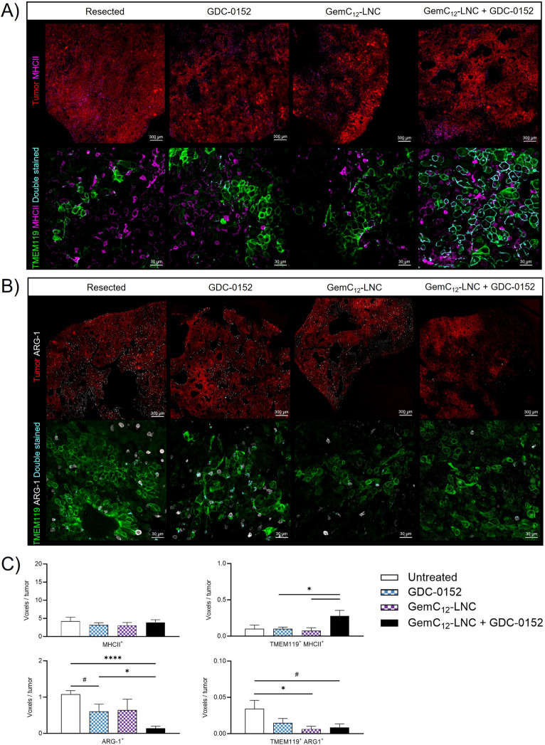
Fig. 8.
A combinatory approach to reverse the post-surgical immunity. A-B) Representative confocal microscopy images of coronal brain slices from animals who had received resection or resection plus treatment(s). Sections were immuno-stained against MHCII (lower panel A, in purple), ARG-1 (lower panel B, in white) and TMEM119 (lower panels A and B, in green) while tumor cells transgenetically expressed DsRed (in red in the upper panels A and B). MHCII+/TMEM119+ cells (panel A) and ARG-1+/TMEM119+ (panel B) are represented in cyan in the lower panels to evaluate the level of microglial activation and the anti-inflammatory status, respectively. Scale bar: 300 µm for upper panels, 30 µm for lower panels; C) Quantifications of the densities of each cell sub-population (mean ± SEM, n = 4; Unpaired t test, # 0.05 < p < 0.06; *p < 0.05; ****p < 0.0001)

