
FIGURE 4.
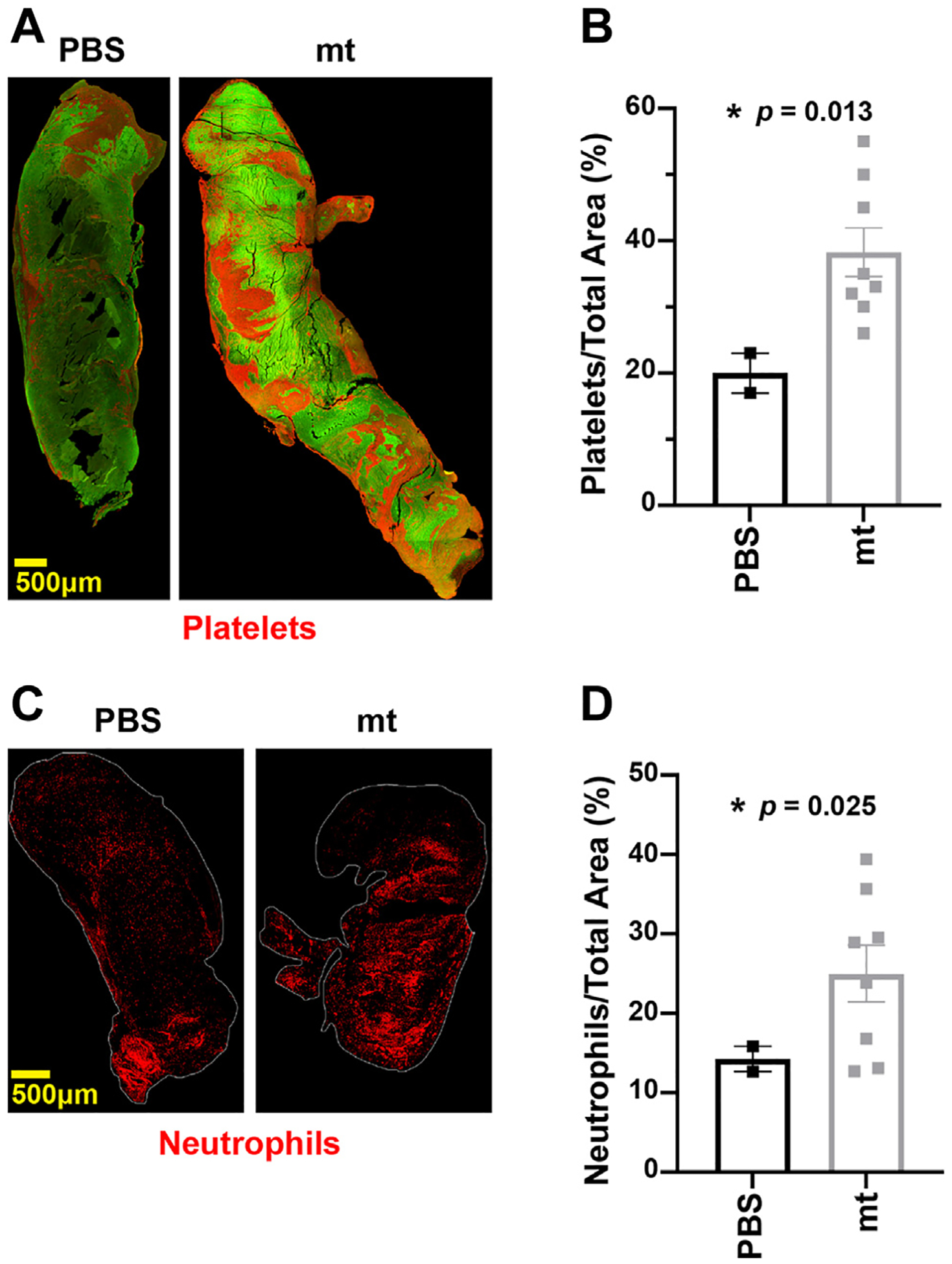
Mitochondria (mt)-induced venous thrombi are rich in platelets and neutrophils. (A) Representative immunofluorescence stain for platelets (CD42b) in PBS- and mt-induced venous thrombi. (B) Average surface area covered by platelets in PBS- and mt-induced venous thrombi. (C) Representative immunofluorescence stain for neutrophils (Lys6G) in phosphate-buffered saline (PBS)- and mt-induced venous thrombi. (D) Average surface area covered by neutrophils in PBS- and mt-induced venous thrombi.