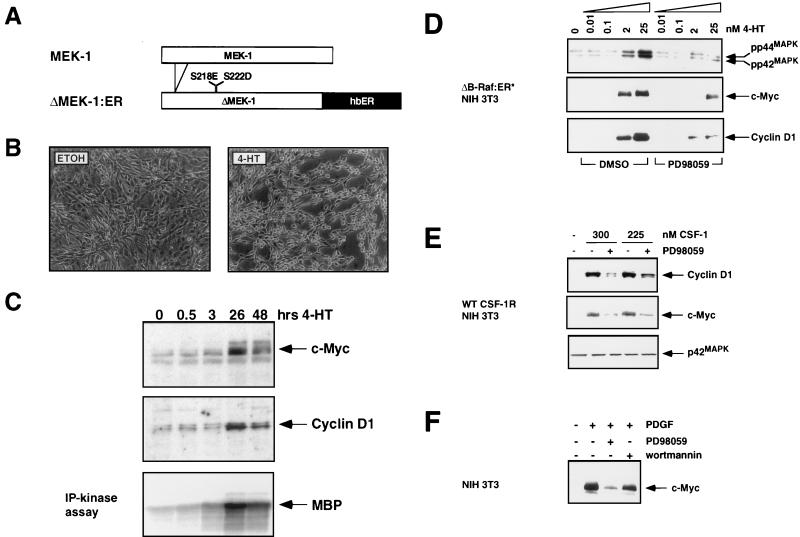
FIG. 5.
MEK activity is sufficient and necessary for the induction of c-Myc and cyclin D1. (A) Construction of ΔMEK1:ER. DNA sequences encoding a constitutively active form of MEK1 (ΔN3, S218E, and S222D) were fused to the hormone-binding domain of the HE14 allele of the human estrogen receptor and cloned into the pBabepuro3 replication-defective retroviral expression vector (64, 72). (B) Morphological transformation of NIH 3T3 cells after activation of ΔMEK1:ER. NIH 3T3 cells expressing ΔMEK1:ER were cultured in DSFM for 24 h and then treated with either ethanol (solvent control) or 1 μM 4-HT for 48 h as indicated, at which time photomicrographs were taken. (C) MAP kinase activity and expression of c-Myc and cyclin D1 after ΔMEK1:ER activation. NIH 3T3 cells expressing ΔMEK1:ER were cultured in DSFM for 24 h and then treated with 1 μM 4-HT for various lengths of time. Expression of c-Myc (top panel) and cyclin D1 (middle panel) was detected by Western blotting, and MAP kinase activity was assessed by p42 and p44 immune complex kinase assay with MBP as a substrate (lower panel). (D) Effect of inhibiting MEK activity on Raf induction of c-Myc and cyclin D1 expression. NIH 3T3 cells expressing ΔB-Raf:ER* were cultured in DSFM for 24 h and then treated with either DMSO (solvent conrol) or 100 μM PD098059 for 40 min. Different concentrations of 4-HT were then added, and cells were harvested after either 6 h (top and middle panels) or 25 h (bottom panel). The activities of the p42 and p44 MAP kinases (top panel) were measured by immune-complex kinase assay, and expression of c-Myc and cyclin D1 (middle and bottom panels) was assessed by Western blotting. (E) Effect of inhibiting MEK activity on CSF-1 induction of c-Myc and cyclin D1. NIH 3T3 cells expressing the wild-type CSF-1 receptor (WT CSF-1R) were cultured in DSFM for 20 h and then treated with either DMSO or 100 μM PD098059 for 40 min. Cells were then treated with 300 or 225 nM CSF-1 and harvested after either 1 h (middle and bottom panels) or 22.5 h (top panel). c-Myc and cyclin D1 expression (top and middle panels) was assessed by Western blotting. The c-Myc blot was reprobed with an anti-p42 MAP kinase antiserum as a control for equal loading. (F) Effect of inhibiting MEK activity on PDGF induction of c-Myc. Parental NIH 3T3 cells were cultured in DSFM for 16 h and then treated with either 100 μM PD098059, 10 nM wortmannin, or DMSO as a solvent control for 40 min. Cells were then treated with 10 ng of PDGF per ml and harvested 2 h later. c-Myc expression was assessed by Western blotting.

