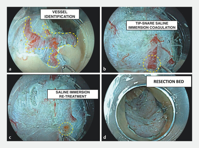
Fig. 1.
Endoscopic images of saline-immersion coagulation. a Blood vessel identification (dashed line) after endoscopic mucosal resection. b Prophylactic snare-tip coagulation. c The vessels appear whitish after application of the high-current voltage under saline immersion. d Resection bed after saline-immersion snare-tip vessel coagulation.

