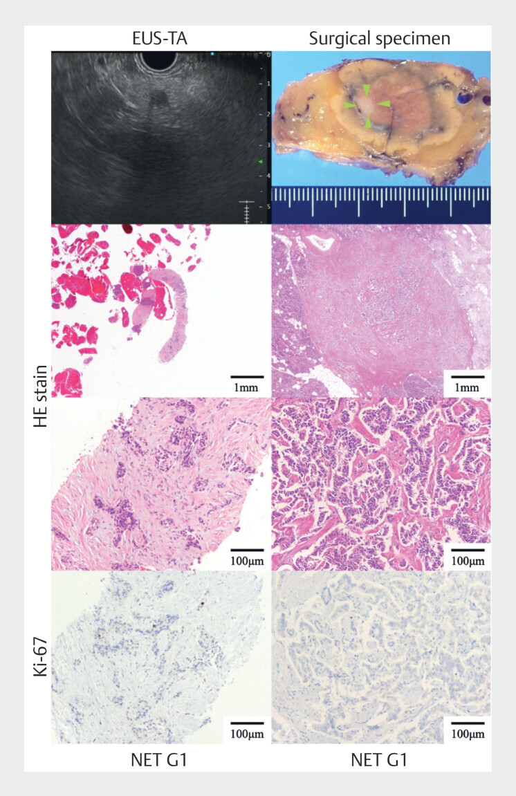
Fig. 2.
Representative images of diagnosis of small pancreatic neuroendocrine neoplasm using endoscopic ultrasound-guided tissue acquisition (EUS-TA). The smallest tumor diameter for which grading in this study could be assessed was 5 mm. The left row shows the EUS-TA specimen and the right row shows the surgical specimens (green arrowheads indicate the tumor site). The uppermost left row shows the EUS image and the uppermost right row shows the surgical specimen. The middle two rows are hematoxylin and eosin-stained sections and the bottom row is a section stained for Ki-67.

