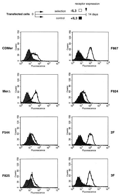
FIG. 3.
Increased receptor expression after selection of cells in the absence of IL-3. Cells expressing the indicated receptors were maintained for 14 days in medium with (+) IL-3 (control cells) (filled-in curve) or without (−) IL-3 (selected surviving cells) (unfilled curve). The expression of the receptors was detected by FACScan analysis using an FITC-conjugated anti-CD8 antibody. A scheme of the selection process is shown above the receptor expression histograms.

