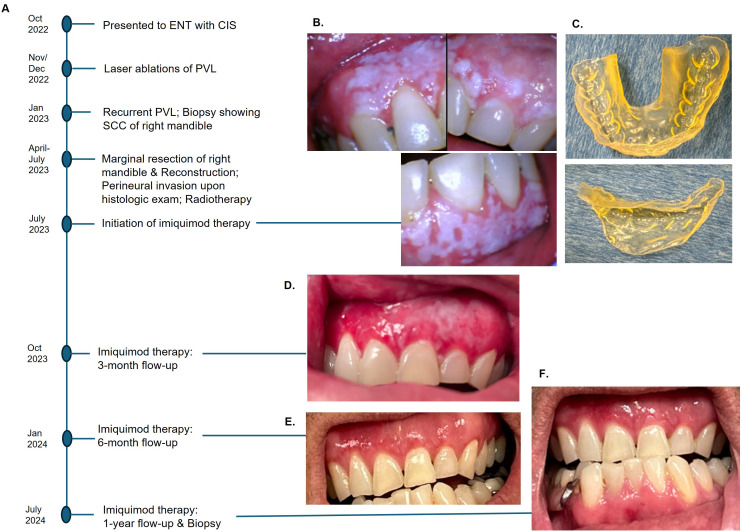
Figure 1.
Events in chronological order (A). Diffuse PVL of maxillary gingiva and left mandibular gingiva after marginal resection of the right posterior mandible (B), 3-D printed customized tray to hold imiquimod cream (C). A significant reduction of PVL was observed at 3-month follow-up (D), and no clinically visible lesion at 6-month post-therapy (E). No clinical or histologic evidence of recurrence after a year with continued use of imiquimod (F).

