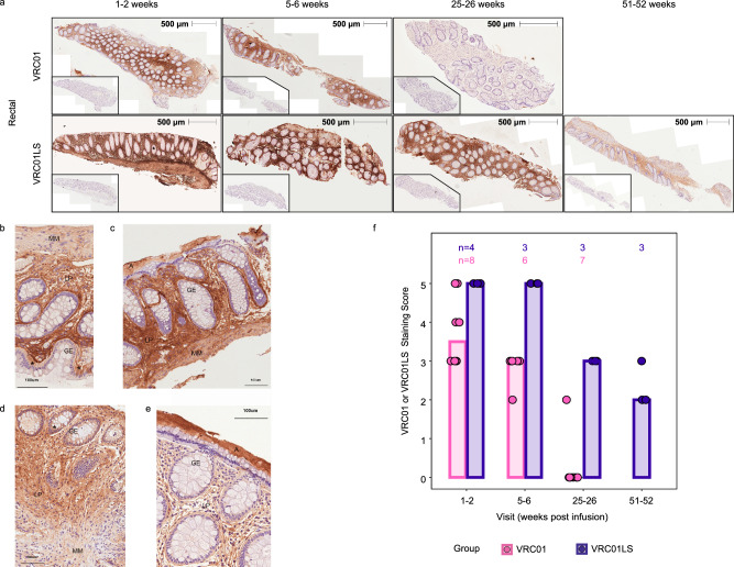
Fig. 4. Infused VRC01 and VRC01LS localizes into the rectal lamina propria and muscularis layers.
PAXgene-fixed, paraffin-embedded rectal tissue biopsies were sectioned (4 μm) and stained with 5C9 (VRC01 and VRC01LS detection) or mouse IgG2a (isotype control), followed by anti-mouse IgG-DAB (brown staining), and counterstained with hematoxylin (blue). All images were acquired on the TissueFAXS microscope in a bright field using a 10 × objective and identical exposure times. a Whole tissue section images of two representative individuals from VRC01-infused (n = 8) and VRC01LS-infused recipients (n = 4) are shown at multiple time points. Whole tissue section images indicate 5C9 staining of VRC01 or VRC01LS with 500 μm measurements to indicate the size of the tissue section analyzed; inset images are consecutive sections to the 5C9 stains, stained with mouse IgG isotype control. b–e Sections from 4 different participants at high magnification, with adjacent isotype control stained images. b, c Sections of rectal biopsies from 2 different VRC01-infused participants at 5, 6 weeks post infusion. d, e Sections of rectal biopsies from 2 different VRC01LS-infused participants at 5, 6 weeks post infusion. Abbreviations: A: adherent mucus layer, GE: glandular epithelium, LP: lamina propria, MM: muscularis mucosa, *: selected epithelium depicting mAb staining. A 100 μm ruler indicates size. f Manual scoring of 5C9 staining at different time points post-infusion. The number of participant samples analyzed is indicated above each bar. Scores were subtracted for any mouse IgG2a background to remove any artifacts. No data was excluded.

