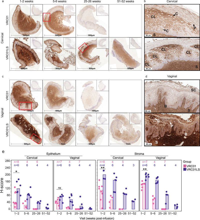
Fig. 5. Localization of VRC01 and VRC01LS in cervical and vaginal tissues after infusion.
a, c Whole tissue section images showing persistence of VRC01LS compared to VRC01 in (a) cervical and (c) vaginal tissues; inset images show isotype control staining on consecutive sections. All time points for each tissue type are from the same representative VRC01 (n = 8) or VRC01LS (n = 6) recipient. b, d Higher magnification views of regions from (a) and (c), outlined in red, showing specific localization patterns of VRC01 or VRC01LS within (b) cervical and (d) vaginal epithelium. Arrows labeled (c) and P indicate examples of cytoplasmic and pericellular localization, respectively. Examples of localization in the stratum corneum and basal (and parabasal) layers are labeled SC and (b), respectively, and stromal staining is labeled S. In panel (b), there is a combination of pericellular, cytoplasmic, and clustered localization (labeled CL) in the intermediate layer in both cervical images; upper and lower images show a single large area and five smaller areas, respectively, of clustered localization in the intermediate layers. In panel (d), the upper vaginal image shows pericellular localization with some concentration in the basal, parabasal, and stratum corneum. The lower image shows pericellular localization and regions of cytoplasmic localization with some concentrated deposition in the intermediate layer as well as the basal, parabasal, and most superficial layers of the stratum corneum. e Area quantitation of VRC01 or VRC01LS staining over time post-infusion in cervical and vaginal epithelium and stroma. The number of participant samples analyzed is indicated above each bar. Individual data points correspond to the H-scores for each participant sample; bar heights represent the medians. Three cervical and 2 vaginal samples had insufficient tissue, and 1 cervical sample had insufficient stroma and were therefore excluded from analysis. Two-sided Wilcoxon rank sum test comparisons were done at 1-2 weeks post-infusion. The exact p-values are as follows: cervical (*) p = 0.020 and vaginal (ns, not significant) p = 0.051 epithelium; cervical (**) p = 0.004 and vaginal (**) p = 0.001 stroma. Of note, VRC01LS recipients donated week 1-2 samples slightly earlier (median 7.5 days, range 4–14) than VRC01 recipients (median 11 days, range 2–15).

