图3.

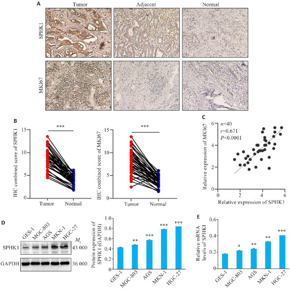
GC组织中SPHK1表达的上调
Fig.3 SPHK1 expression is upregulated in GC tissue. A: Immunohistochemical staining for detecting SPHK1 and MKI67 in clinical tissue samples (Original magnification:×200). B: Immunohistochemical score of SPHK1 and MKI67. ***P<0.001. C: Correlation between SPHK1 and MKI67 expressions. D: Western blotting for detecting the expression of SPHK1 protein. E: qRT-PCR for detecting the level of SPHK1 mRNA. *P<0.05, **P<0.01, ***P<0.001 vs GES-1 group.
