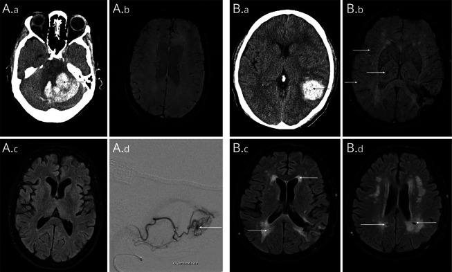
Figure 3. Neuroimaging Examples Showing Included Patients With Different Risks of a Macrovascular Cause.
(A) Patient in their fifties with cerebellar ICH (A.a). SWI and FLAIR sequences (A.b/A.c) showed no evidence of cerebral small vessel disease (MACRO score 1, indicating a high risk of a macrovascular cause). In dynamic MR angiography, a small AVM originating from the left posterior inferior cerebellar artery was suspected, which was confirmed on DSA (A.d). (B) Patient in their forties with a parietal lobar ICH (B.a). SWI shows several cerebral microbleeds in a mixed distribution (B.b), as well as confluent white matter hyperintensities and several lacunes (B.c/B.d). This patient had a MACRO score of 7, indicating a very low risk of a macrovascular cause. DSA and repeat MRI did not show any evidence of a macrovascular cause for the ICH. DSA = digital subtraction angiography; FLAIR = fluid-attenuated inversion recovery; ICH = intracerebral hemorrhage; MACRO = MRI Assessment of the Causes of intRacerebral haemOrrhage.

