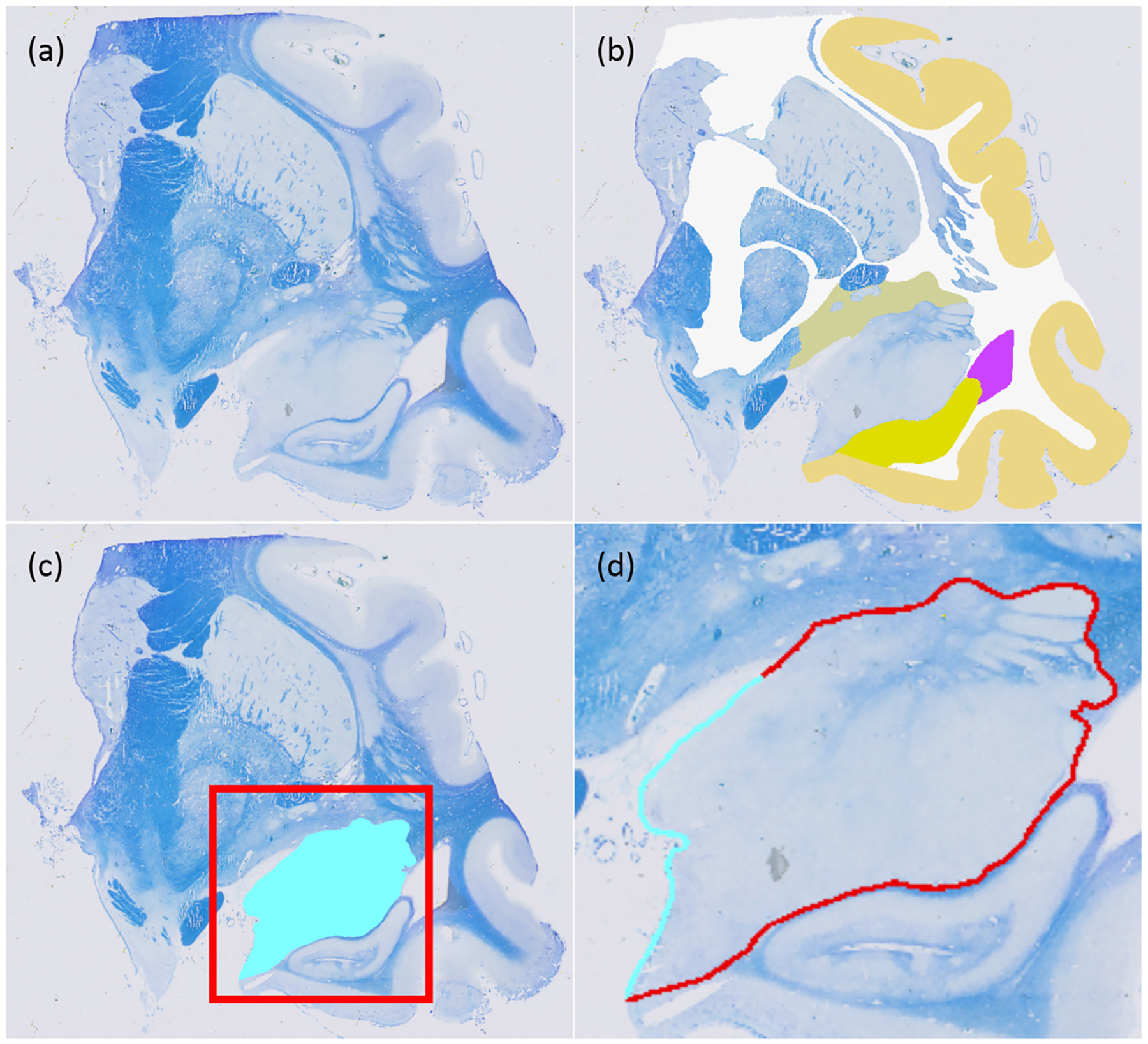
Fig. 2.

(a) Sample histological section. (b) Available manual annotations at iteration of the active learning process. (c) Query at : the algorithm has requested labelling of the amygdala on this section (in light blue). (d) The actual boundary length that needs to be labelled (in light blue) is much smaller than the perimeter of the amygdala, since most of it has already been labelled as part of neighbouring structures (in red).
