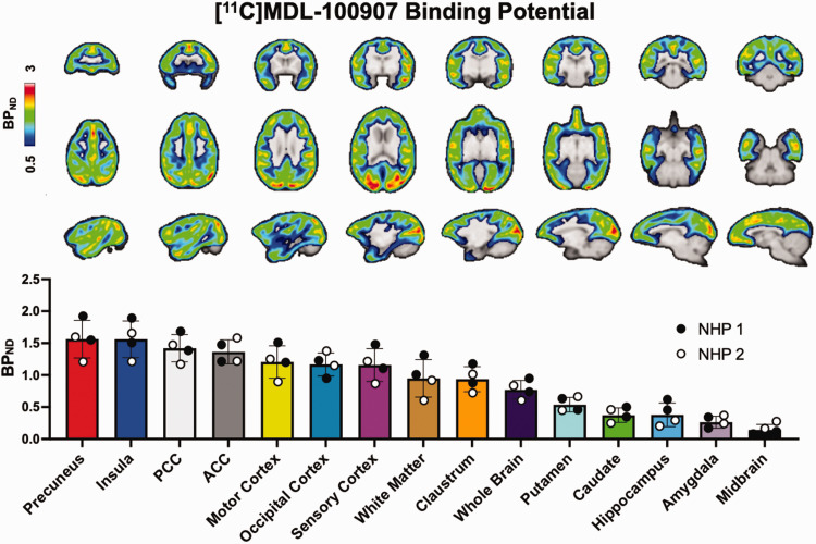
Figure 1.
Baseline BPND of [11C]MDL-100907 in non-human primate (NHP) brains. Representative images are in axial, coronal, and sagittal view. Region-specific BPND are shown from in descending order from highest to lowest observed binding for the ROI sampled. White and black dots represent NHP-specific datapoints.

