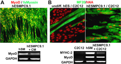
Figure 3. Myogenic Differentiation of hESMPCs.
(A) Immunocytochemistry for MyoD (red) and fast-switch myosin (green). RT-PCR for MyoD in human skeletal muscle as a positive control (hSM), and in hESMPC-H9.1 cells differentiated for 10 d in the presence of C2C12-conditioned medium (hESMPC).
(B) Myotube formation induced at high cell densities in the presence of C2C12 cells. Myotube characterization by immunocytochemistry for MF20 against sarcomeric myosin (green) and human nuclear antigen (hNA, red). Left panel: Control undifferentiated hESCs (H9) do not fuse with C2C12. Right panel: Under identical culture conditions, hESMPCs (line 9.1) efficiently fuse with C2C12 cells, forming myotubes containing human nuclei. RT-PCR for human specific muscle transcripts myosin heavy chain IIa (MYHC-2) and MyoD in C2C12 cells, in human skeletal muscle as positive control (huSM), and in hESMPC-H9.1 cells cocultured with C2C12 cells.

