Fig. 6.
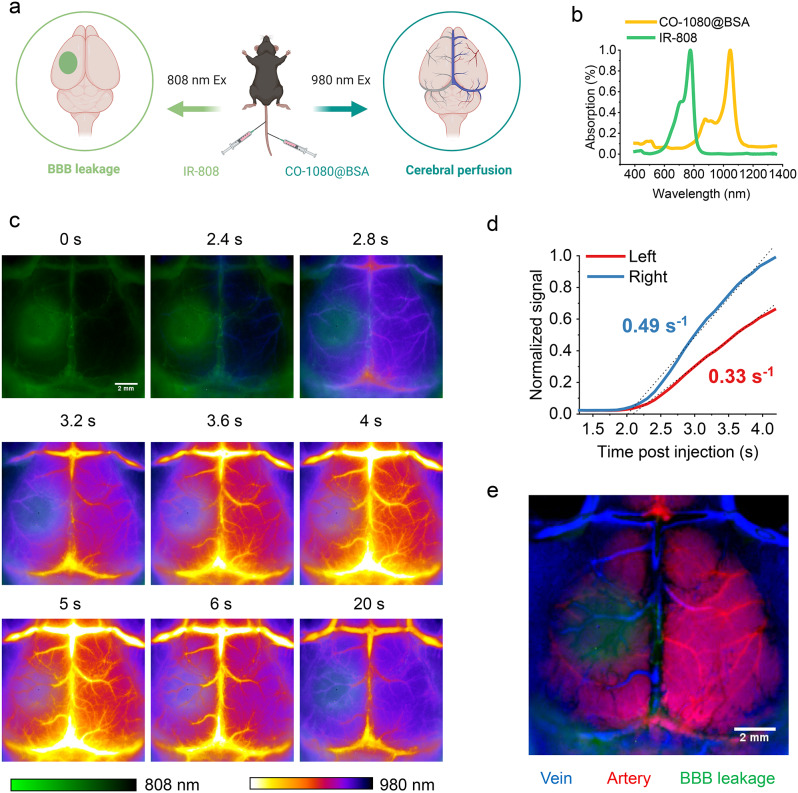
Dual-channel NIR-II imaging of BBB disruption and cerebral perfusion after stroke. (a) Schematic diagram of dual-channel for BBB and cerebral perfusion imaging (Created with BioRender.com). (b) The absorption spectra of IR-808 and CO-1080@BSA. (c) Dual-channel images of the BBB and cerebral vascular (100 ms, 1200 LP) after stroke at time post CO-1080@BSA injection. IR-808 was used to evaluate BBB disruption using 808 nm laser excitation, while CO-1080@BSA was used to dynamically evaluate the cerebral vascular using 980 nm excitation at 6 h post stroke. (d) Fluorescence intensity (normalized to the right-side peak) versus time in the left (infarct side) and right (contralateral) sides of the brain of mice with stroke showed a significant reduction in perfusion in the stroke area. (e) Dynamic sequential images of vessels with CO-1080@BSA were analyzed by principal component analysis and merged with images of the BBB leakage area (green) to resolve arterial (red) and venous (blue) cerebral vessels

