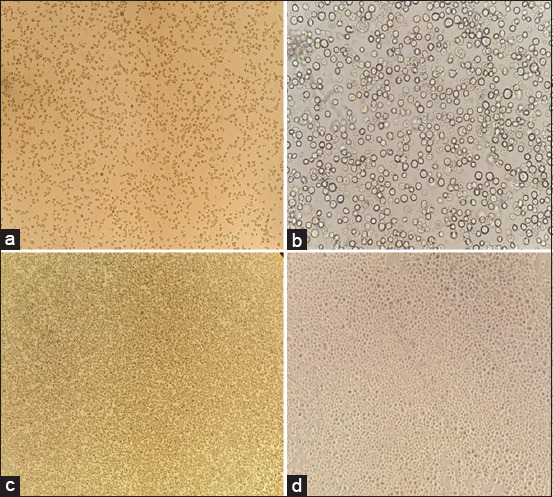
Figure-4.

Monolayer of SF9 cells prepared at different time points. SF9 cells show confluency at different time periods, (a) after the addition of cells from the storage vial, most cells were suspended, (b) after 24 h the confluency of the cells at 10×, (c) cells confluency (80%–85%) after 48–60 h, (d) 100% confluency of the SF9 cells.
