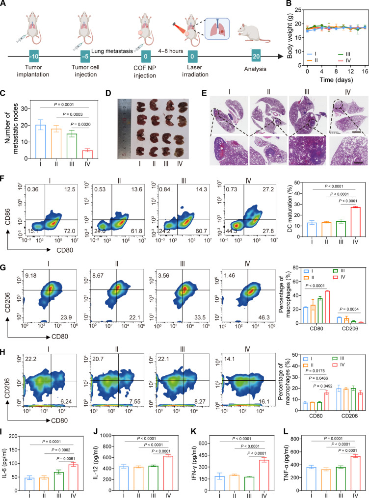
Fig. 8. Therapeutic effect of TPAD-COF NPs on a pulmonary metastasis model under NIR laser irradiation.
(A) Scheme of lung metastasis model establishment and treatment process. (B) Mouse body weights (n = 5), (C) number of metastatic nodes in lung tissues (n = 4), (D) digital photograph of the dissected lung tissues (n = 4), and (E) H&E staining images of the lung tissues after different treatments. (F) Flow cytometry analysis and corresponding statistical analysis of DC maturation in tumor tissues of each group (n = 3). (G and H) Flow cytometry analysis of M1 and M2 macrophage proportions in (G) tumor tissues and (H) spleen tissues after different treatments and the corresponding statistical analysis (n = 3). (I to L) The secreted levels of cytokines (n = 3), including (I) IL-6, (J) IL-12, (K) INF-γ, and (L) TNF-α after diverse treatments [I: Control; II: Laser (1.0 W cm−2, 10 min); III: TPAD-COF NPs (100 μg ml−1); IV: TPAD-COF NPs (100 μg ml−1) + laser (1.0 W cm−2, 10 min)].

