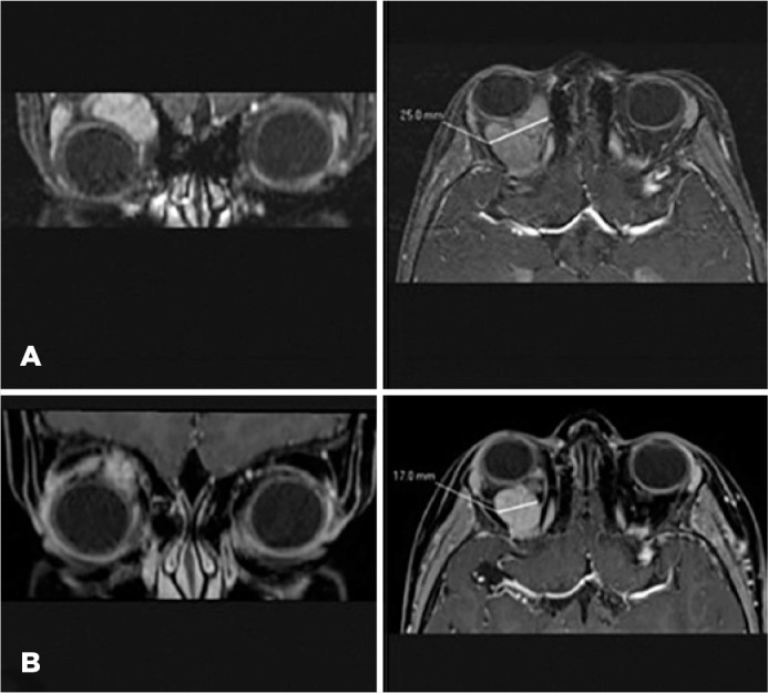
Figure 2.

Cranial MRI, orbital section. Coronal (left) and axial (right) postcontrast SPIR T1WI. (A) Well defined and avid enhancing mass located in the right orbital space, consistent with orbital venous-lymphatic malformation. The lesion extends into the intraconal fat with shifting in associated structures resulting in proptosis (right) and infero-medial deviation of the right eye (left). There was no local infiltration observed. (B) Decrease in proptosis (left) and mass size from 25 mm (Figure 2A, right) to 17 mm (Figure 2B, right) after 3 months of treatment with sirolimus.
MRI, magnetic resonance imaging; SPIR, spectral presaturation with inversion recovery
