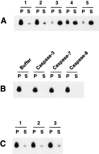
FIG. 5.
Cif activation by caspases in the S-100 cytosolic extract from Bcl-2:HL-60 cells. (A) Aliquots (100 μg) of S-100 cytosolic extract from Bcl-2:HL-60 cells were left untreated (lane 1) or treated with 5 μl of buffer alone (lane 2), 5 μl (100 ng) of caspase 3 (lane 3), 5 μl (100 ng) of caspase 7 (lane 4), or 5 μl (100 ng) of caspase 8 (lane 5) for 60 min at 37°C. The samples were then incubated with HL-60 mitochondria for 15 min at 37°C and then centrifuged to pellet the mitochondria. The pellet (P) and supernatant (S) were separated, and the cytochrome c content in each fraction was determined by Western blot analysis. (B) HL-60 mitochondria were incubated with buffer alone or 100 ng of caspase 3, caspase 7, or caspase 8 for 15 min at 37°C and then analyzed as described for panel A. (C) Aliquots (100 μg) of S-100 cytosolic extract from Bcl-2:HL-60 cells were treated with 100 ng of caspase 3 (lane 1), caspase 7 (lane 2), or caspase 8 (lane 3) for 60 min at 37°C. The samples were then heated at 100°C for 10 min, incubated with mitochondria for 15 min at 37°C, and assayed as described for panel A.

