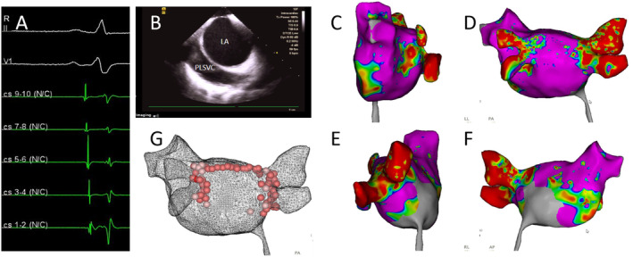
FIGURE 1.

(A) The preoperative electrocardiogram indicates sinus rhythm. (B) Intracardiac echocardiography (ICE) shows an echolucent shadow alongside the left atrium (LA), consistent with a persistent left superior vena cava (PLSVC). (C) Three‐dimensional electroanatomical map of the left atrium in sinus rhythm, viewed from the left side. (D) Three‐dimensional electroanatomical map of the left atrium in sinus rhythm, viewed from the posterior–anterior perspective. (E) Three‐dimensional electroanatomical map of the left atrium in sinus rhythm, viewed from the right side. (F) Three‐dimensional electroanatomical map of the left atrium in sinus rhythm, viewed from the anterior–posterior perspective. (G) Ablation steps include gap ablation within the pulmonary veins, expansion of the ablation area, and linear ablation at the roof.
