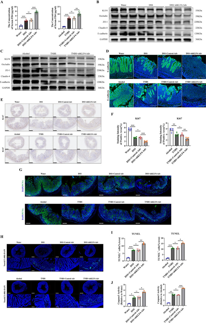
Fig. 3.
KLF4 downregulation impairs intestinal epithelial barrier integrity by decreasing AJC protein expression and increasing apoptosis of IECs in DSS- and TNBS-induced colitis. A The serum concentration of FD4 in DSS- and TNBS-induced colitis mice after treatment with shKLF4-Adv. WT + water (n = 6), WT + DSS (n = 8), control-Adv + DSS (n = 5), or shKLF4-Adv + DSS (n = 10); WT + alcohol (n = 6), WT + TNBS (n = 8), control-Adv + TNBS (n = 5), or shKLF4-Adv + TNBS (n = 10). B, C The protein levels of KLF4, E-cadherin, Occludin, ZO-1 and claudin-4 in colon tissues from DSS (B)- and TNBS (C)-challenged mice after treatment with shKLF4-Adv were assessed by Western blot. D Representative immunofluorescence images of E-cadherin (green) in colon tissues from DSS- and TNBS-challenged mice after treatment with shKLF4-Adv. Scale bars, 100 μm. E, F The expression of Ki67 in colon tissues from DSS- and TNBS-challenged mice after treatment with shKLF4-Adv was detected by IHC. Scale bars, 500 μm (top) and 250 μm (bottom). The staining intensity of KLF4 based on the IHC staining index. G Representative immunofluorescence images of PCNA (green) in colon tissues from DSS- and TNBS-challenged mice after treatment with shKLF4-Adv. Scale bars, 200 μm. H Representative TUNEL images of colon tissues from DSS- and TNBS-challenged mice after treatment with shKLF4-Adv. Scale bars, 700 μm (top) and 100 μm (bottom). I Quantitative analysis of TUNEL-positive cells in colon tissues from DSS- and TNBS-challenged mice after treatment with shKLF4-Adv. J Quantitative analysis of Caspase3 activity in colon tissues from DSS- and TNBS-challenged mice after treatment with shKLF4-Adv. Data are expressed as the mean ± SD. ns, not significant, * p < 0.05, ** p < 0.01, *** p < 0.001

