Fig. 4.
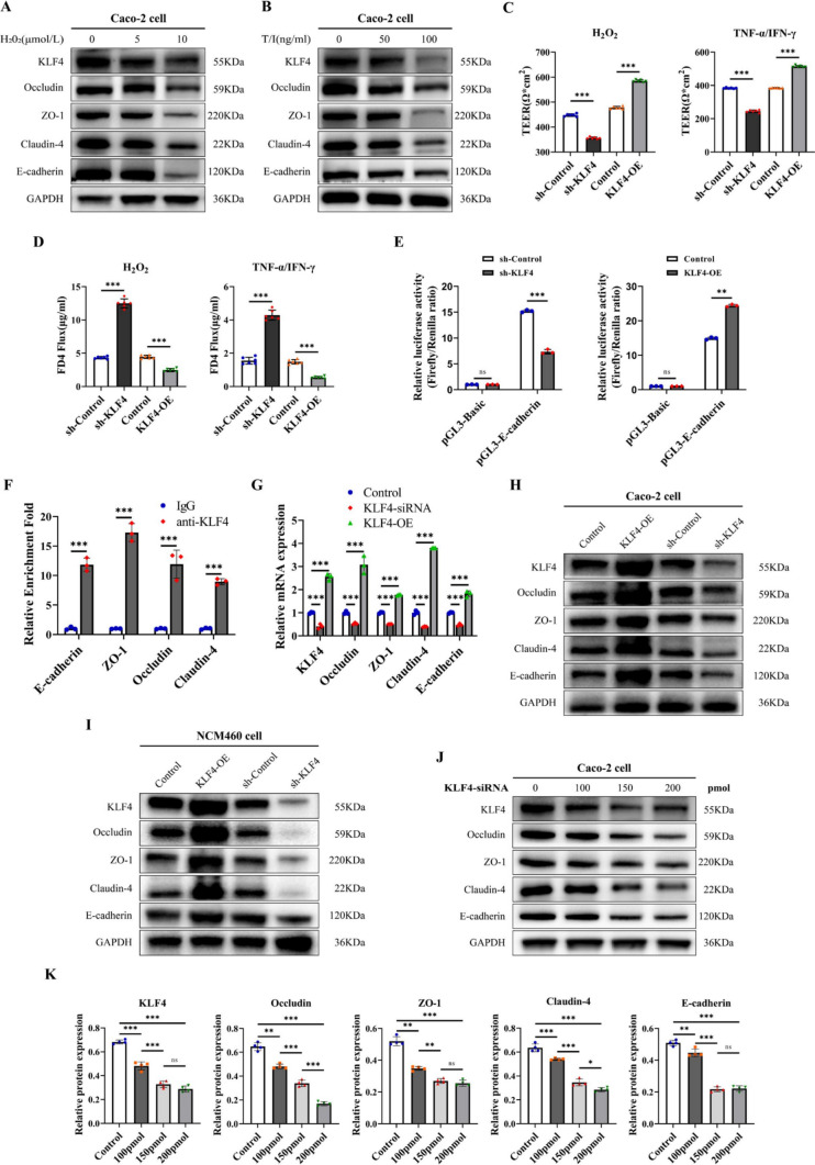
KLF4 knockdown leads to intestinal epithelial barrier damage by regulating AJC proteins in vitro. A, B KLF4, Occludin, ZO-1, Claudin-4 and E-cadherin protein expression in Caco-2 cells after treatment with different doses of H2O2 (A) or TNF-α/IFN-γ (B). C, D TEER (C) and FD4 permeability (D) were measured in KLF4-overexpressing or KLF4-knockdown Caco-2 cells after stimulation with H2O2 (10 μmol/L) and TNF-α/IFN-γ (100 ng/ml). E The luciferase activity, responsive to KLF4, was measured in KLF4-overexpressing or KLF4-knockdown Caco-2 cells co-transfected with renilla luciferase plasmids and apGL3-Basic-Luciferase reporter vectors containing the promoter sequence of E-cadherin. F ChIP analysis of KLF4 binding to the promoters of E-cadherin, Occludin, ZO-1, and Claudin-4 in Caco-2 cells. The relative enrichment was calculated as the fold change in DNA abundance following pull-down of anti-KLF4 or normal rabbit IgG. G The mRNA expression levels of KLF4, Occludin, ZO-1, Claudin-4 and E-cadherin in KLF4-knockdown or KLF4-overexpressing Caco-2 cells were examined by RT‒qPCR. H, I The protein expression of KLF4, Occludin, ZO-1, Claudin-4 and E-cadherin in Caco-2 cells (H) and NCM460 cells (I) infected with sh-KLF4 lentivirus and KLF4-overexpressing lentivirus. J The protein expression of KLF4, Occludin, ZO-1, Claudin-4 and E-cadherin in Caco-2 cells treated with siKLF4 at 100 pmol, 150 pmol, or 200 pmol. K The Western blot band densities shown in Fig. 4 J were quantified using ImageJ software. The data are expressed as the mean ± SD. ns, not significant, * p < 0.05, ** p < 0.01, *** p < 0.001

