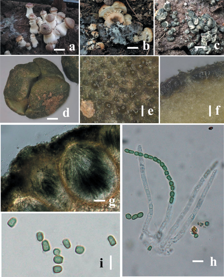
Figure 3.
Morphology of Trichodermastrophariensis (HGUP 24-0001, GUCC 24-0002) a, b disease in the field habitat c fresh stromata on natural habitat d dry stromata e ostiolar dots on stromata surface f cortical and subcortical tissues in section g ascomatal tissue in section h asci with ascospores i ascospores. Scale bars: 10 mm (a, b); 20 mm (c); 100 mm (d–f); 50 μm (g); 20 μm (h, i).

