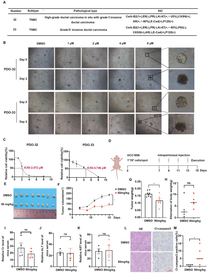
Fig. 6.
YZ-836P inhibits growth of TNBC patient-derived organoids and xenograft tumors in vivo. (A) Pathological data of two TNBC patients. (B) YZ-836P disrupted the structural integrity of PDOs. Microscopic observation of PDOs morphology before and after 48 h of YZ-836P treatment. Arrows indicated decomposing PDOs. (C) YZ-836P inhibited ATP activity in PDOs. Cell viability changes in PDOs treated with YZ-836P for 48 h, measured by using an ATPase activity assay kit. (D) Schematic of the xenograft tumor model in nude mice and treatment protocol. (E) YZ-836P inhibited xenograft tumor growth. Mice were sacrificed 15 days after tumor inoculation, and tumors were collected and photographed (n = 8 per group). (F) YZ-836P reduced xenograft tumor volume. Tumor volume was measured every other day starting from the 7th day after inoculation (n = 8 per group). (G) YZ-836P decreased xenograft tumor weight. Tumor weight was measured after collection (n = 8 per group). (H) Body weight changes in nude mice before and after YZ-836P treatment (n = 4 per group). (I) YZ-836P treatment did not significantly affect serum creatinine (Cr) levels (n = 4 per group). (J) YZ-836P treatment did not significantly affect serum alanine aminotransferase (ALT) levels (n = 4 per group). (K) YZ-836P treatment did not significantly affect serum aspartate aminotransferase (AST) levels (n = 4 per group). (L) Immunohistochemical analysis of cl-Caspase 3 expression in xenograft tumors, with representative images. (M) YZ-836P treatment significantly increased cl-Caspase 3 expression in xenograft tumors (n = 6 per group). Data represent results from three independent experiments. *p < 0.05, **p < 0.01, and ***p < 0.001; ns, not significant

