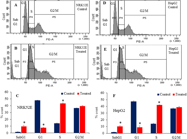
Figure 13.
Cell cycle analysis of NRK52E and HepG2 cells following exposure to 100 µg/mL of Bi2O3/RGO nanocomposites for 48 h. Histogram of control (A), treated (B), and the percentage of cell population in subG1, G1, S, and G2/M phases (C) of NRK52E cells. Histogram of control (D), treated (E), and the percentage of cell population in subG1, G1, S, and G2/M phases (F) of HepG2 cells. Quantitative data is represented as the mean±SD of three separate experiments (n=3). *Significantly different as compared to the control (p<0.05).

