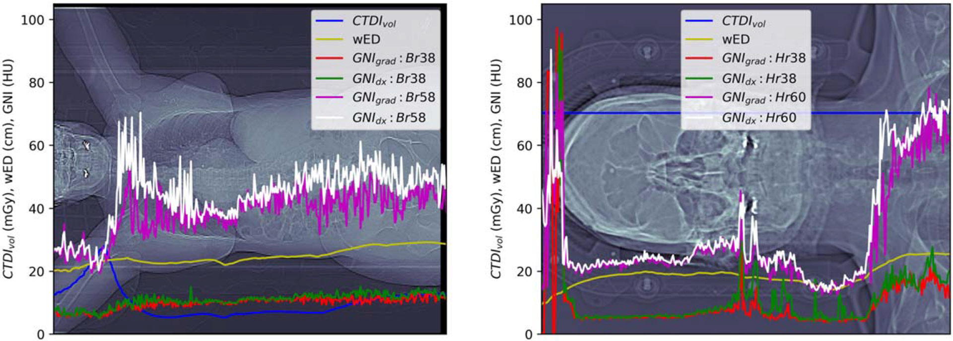
Figure 6.

GNIs computed at each CT slice reconstructed with regular (Br38 or Hr38) or sharper (Br58 or Hr60) kernels. CTDIvol and wED was the scanned CTDIvol extracted from dicom of the image slice and the calculated wED. The wED at the shoulder of the brain scan was underestimated due to the CT cut off of the shoulders due to the small FOV.
