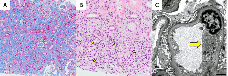
Fig. 2.
Renal biopsy findings are depicted. A Light microscopy revealing interstitial fibrosis and tubular atrophy, using the Masson trichrome staining, (magnification × 200; scale bar = 50 µm). B Intense tubulitis with neutrophilia and lymphocytosis is observed in a few non-atrophic tubular areas, using the periodic acid–Schiff staining (as indicated by the yellow arrowheads; magnification × 400; scale bar = 100 µm). C Electron microscopy revealing mild swelling of the glomerular endothelial cells (as indicated by the yellow arrow; scale bar = 2 µm)

