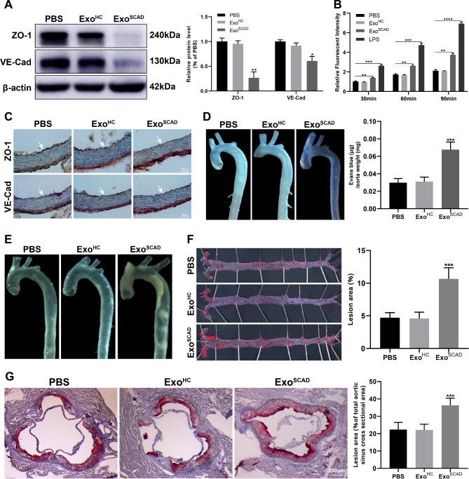
Fig. 3.
ExoSCAD suppress VE-Cad and ZO-1 expressions in ECs and aggravates atherosclerotic lesions. (A) Western blot analyses of abundance of VE-Cad and ZO-1 in HAECs treated with ExoSCAD, ExoHC or PBS. Representative blots (left panel) and the corresponding quantification results (right panel) of 3 replicated independent experiments. (B) Quantification of endothelial permeability by calculating the amount of rhodamine-dextran passing through the monolayer of HAECs treated with ExoSCAD, ExoHC or PBS (n = 3 per group). LPS was used as a positive control. (C) Immunohistochemistry analyses of VE-Cad and ZO-1 expression in aortas from the 12-week HFD-fed ApoE−/− mice injected with ExoSCAD, ExoHC or PBS (n = 6–10 per group). White arrows indicate tunica intima. (D) Representative images of aortas from the 12-week HFD-fed ApoE−/− mice received different treatment after intravenous injection of Evans blue in each group (left panel). Quantitation of vascular permeability by calculating the amount of Evans blue extravasated per milligram of artery in each group (n = 7–9, right panel). (E) Representative bright field images of aortas from the 12-week HFD-fed ApoE−/− mice treated with ExoSCAD, ExoHC or PBS. (F) Representative images of the en face Oil Red O-stained aortas from the 12-week HFD-fed ApoE−/− mice in each group (left panel). Quantification of atherosclerotic lesion area as the percentage of positive stained areas to the respective whole arterial areas (n = 7–9, right panel). (G) Representative microphotographs (left panel) and quantification (right panel) of Oil Red O-stained aortic root sections from the 12-week HFD-fed ApoE−/− mice in each group (n = 7–9). Results are represented by mean ± SEM with 7–9 replicates for each group. ExoHC: plasma exosomes of healthy controls; ExoSCAD: plasma exosomes of patients with stable coronary artery disease; HAECs: human aortic endothelial cells; HFD: high-fat diet; LPS: lipopolysaccharide; ApoE: apolipoprotein E. *p < 0.05, **p < 0.01, ***p < 0.001.

