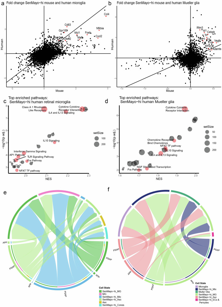
Fig. 5. Comparison of SenMayo-hi glia in mouse and human retina datasets shows conserved interleukin signalling in all glial cells and AP1 pathway enrichment in microglia specifically.
a Scatter plot comparing log fold change of genes in SenMayo-hi microglia (Mic) (against SenMayo-low Mic) in mouse and human retina. b Scatter plot comparing log fold change of genes in SenMayo-hi Mueller glia (MG) in mouse and human retina (against SenMayo-low Mueller glia). c Dotplot of enriched pathways in human SenMayo-hi Mic with pathways that are also enriched in mouse Mic highlighted and labelled in red (FDR < 0.01) See Supplementary Table 9a for a full list. d Dotplot of enriched pathways in human SenMayo-hi MG with pathways that are also enriched in mouse MG highlighted and labelled in red (FDR < 0.01) See Supplementary Table 9b for a full list. e and f Selected common interaction pathways in mouse and human SenMayo-hi cells include APP and PSAP pathways. In both e mouse and f human many interactions of SenMayo-hi cells include signalling to other SenMayo-hi glia.

