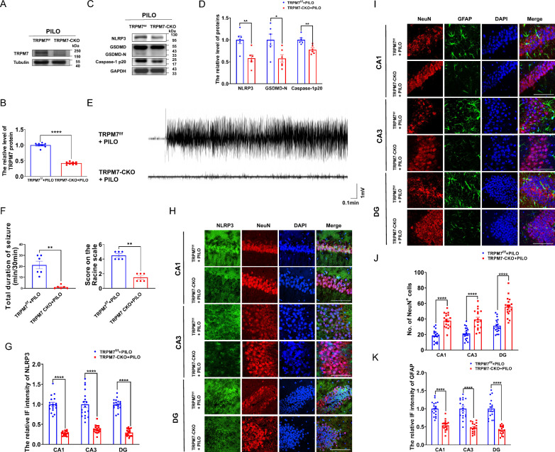
Fig. 9.
TRPM7 conditional knockout mice reduced neuronal damage and pyroptosis and reversed PILO-treated neuronal hyperexcitability. A, B The representative protein bands and analysis of TRPM7 in hippocampus of TRPM7-CKO mice (n = 6). C, D The representative protein bands and analysis of NLRP3, GSDMD, and caspase-1 p20 in hippocampus of TRPM7-CKO mice (n = 6). E Representative 5 min EEG recording showing seizure onset. The TRPM7-CKO mice were injected with PILO. F Left: The mean total duration of seizure activity in PILO-treated TRPM7-CKO mice (n = 6). Right: The score on the Racine scale. G, H Immunofluorescence analysis of NLRP3 (green) and NeuN (red) expression in hippocampus of PILO-treated C57BL/6 J mice (100 × lens), including the CA1, CA3 and DG regions (n = 18). Scale bar: 100 μm. DAPI (blue) was used to label nucleus. I-K Immunofluorescence analysis of NeuN (red) and GFAP (green) expression in hippocampus (100 × lens) of PILO-treated TRPM7-CKO mice, including the CA1, CA3, and DG regions (n = 18). Scale bar: 100 μm. DAPI (blue) was used to label nucleus. Full scans of all the blots are in the Supplementary Note. * P < 0.05; ** P < 0.01; **** P < 0.0001

