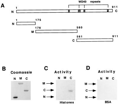
FIG. 5.
The C-terminal region of TFIIICβ harbors the HAT domain. (A) Diagrammatic representation of TFIIICβ (41). Hatched areas indicate positions of WD40 repeats. The lower panel shows TFIIICβ deletion mutants N, M, and C. (B) Coomassie blue stain of equimolar amounts of the His6-tagged TFIIICβ deletion mutants after SDS-PAGE (10% gel). (C and D) In-gel HAT activity assays with purified mutants. TFIIIC mutants were resolved on SDS–10% polyacrylamide gels containing either histones (0.1 mg/ml) (C) or BSA (0.1 mg/ml) as a control (D).

