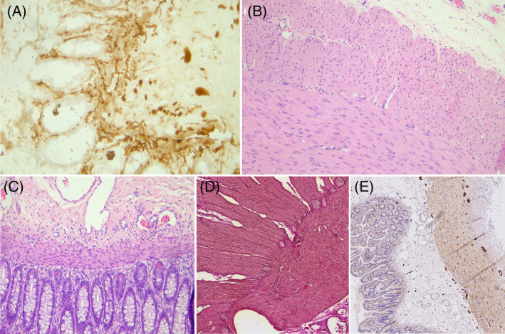
FIGURE 2.

Histopathological examination. Patient 1# (A) Acetylcholinesterase: Initial biopsy comprising rectal mucosa and submucosa where an increase in acetylcholinesterase activity is recognized in the parasympathetic nerve fibers, ganglion cells are not recognizable. (B) Hematoxylin Eosin: Intestinal resection demonstrating a segment with aganglionosis at the level of the myenteric plexus. (C) Hematoxylin Eosin: Intestinal resection demonstrating a segment with aganglionosis at the level of the submucosal plexus. Patient 2#. (D) Elastica‐van‐Gieson staining: Biopsies of the small and large intestine showing ganglia, indicative of hypoganglionosis. (E) S100‐staining: Second biopsy confirming the diagnosis of hypoganglionosis, characterized by increased distances between ganglia.
