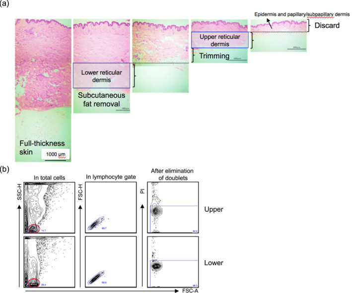
FIGURE 1.

(a) Preparation of the upper and lower reticular dermis (haematoxylin‐eosin staining). The original magnification is ×100. (b) Gating strategy for flow cytometry. In total emigrants, the lymphocyte population indicated that a red circle was gated, followed by the elimination of doublets and dead cells. FSC, forward scatter; PI, propidium iodide; SSC, side scatter.
