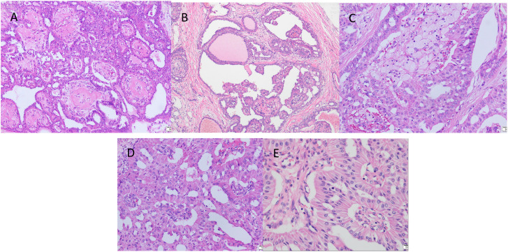
Figure 3.
The postoperative pathological examination with haematoxylin and eosin (HE) staining. (A) Cancer cells arranged in a papillary structure (×100). (B) Cancer cells are arranged in a glandular structure, some of which are cystically dilated, similar to thyroid follicles, with individual glandular lumens containing gelatinous, homogeneous, strongly eosinophilic material, and interstitial fibrous connective tissue (×100). (C) Foam cell aggregates are seen in the interstitium of cancerous tissue (×200). (D) The surface of the papilla is covered with cancer cells in a cubic or high columnar shape, the nucleus is far from the base, the cytoplasm is reddish stained, and the interstitium is seen as a fibrovascular axis (×200). (E) Cancer cells are arranged in high columnar shape with mild nuclear morphology, eosinophilic granular cytoplasm, and nuclei are located away from the base at the luminal margin (×400).

