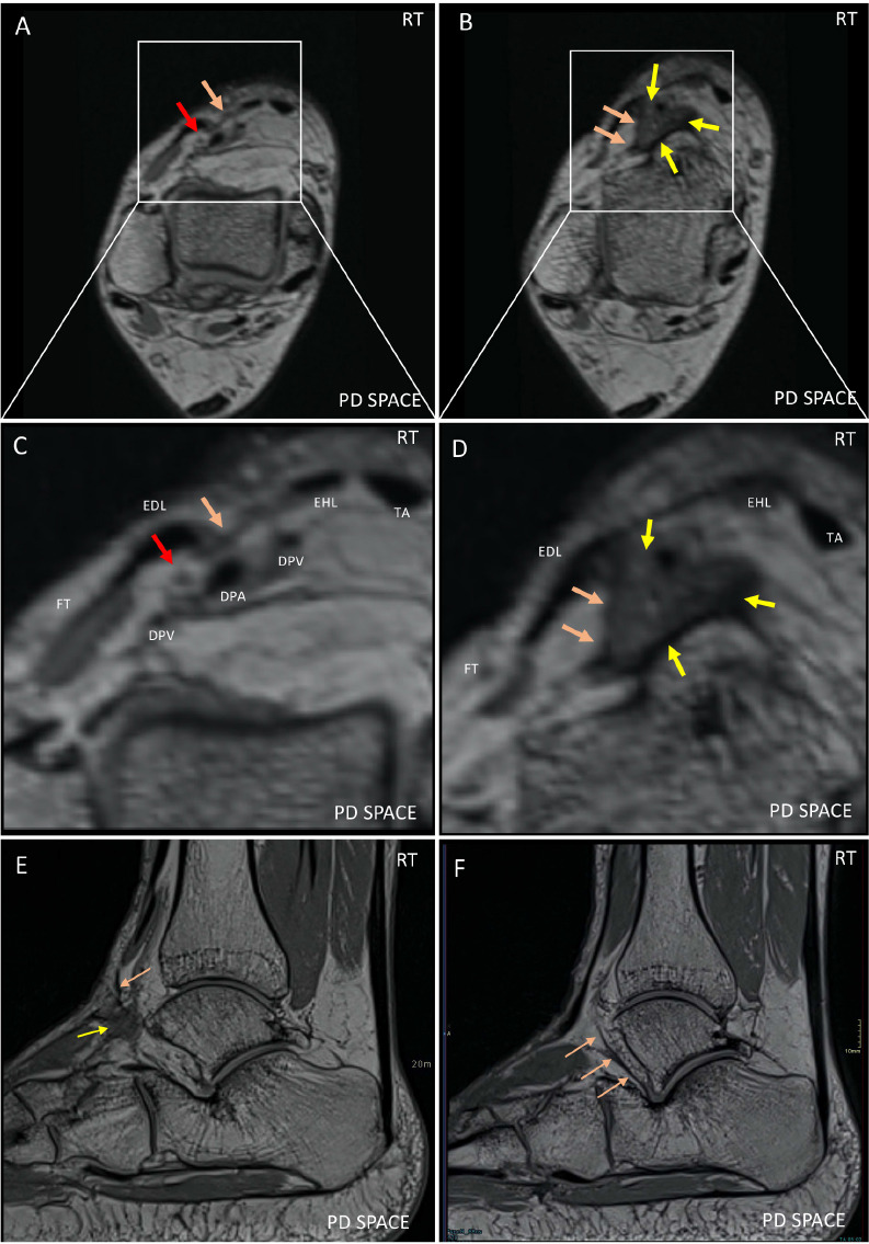
Figure 4.

Axial reformats from PD-weighted 3D sequences of the right ankle above (A) and at the level of the IER (B) with additional magnified views (C, D) as well as the original sagittal images (E, F). C) highlights the arrangement of the deep peroneal neurovascular bundle, also containing the dorsalis pedis artery (DPA) and veins (DPV), proximal to the ATT. The deep peroneal nerve (DPN) is slightly superficial to the DPA and DPVs, indicated by the red arrow. The tibialis anterior (TA), extensor digitorum longus (EDL), extensor hallucis longus (EHL), and peroneus tertius tendons (PT) are located more superficially, as is the IER (orange arrow). D) highlights the geographic region of signal alteration (yellow arrow) encasing the DP neurovascular bundle, at the level of the ATT. The lateral border of the scarring is bordered by the deep lamina (orange arrows) of the IER. Sagittal PD-weighted 3D images of the right ankle (E, F) show the geographic area of signal alteration (yellow arrow) with lateral margin contacting the deep lamina of the inferior extensor retinaculum (orange arrows).
