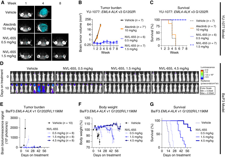
Figure 5.
NVL-655 inhibits intracranial ALK-driven tumor xenografts in mice. A–D, NVL-655 inhibited tumor growth in the YU-1077 intracranial model. A, Superimposed MRI images from individual mice with brain tumor masses highlighted in cyan. B, Brain tumor volume over time based on MRI reconstruction, plotted as mean ± SEM. Horizontal gray line indicates initial tumor volume of the vehicle group. * indicates that six mice remained at the final treatment timepoint in the alectinib treatment group. C, Survival analysis. D–G, NVL-655 inhibited the Ba/F3 EML4–ALK v1 G1202R/L1196M luciferase intracranial model. D, Bioluminescence images indicating a change in tumor burden over 10 days of treatment. E, Brain bioluminescence over time plotted as mean ± SEM. Plotting for each treatment group stopped when the first animal was lost. Horizontal gray line indicates initial bioluminescence of the vehicle group. F, Body weight over time plotted as mean ± SEM. Horizontal gray line indicates initial body weight. G, Survival analysis. All treatment was administered orally twice daily, except alectinib 10 mg/kg, which was administered orally once daily.

