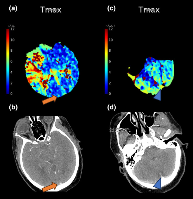
FIGURE 2.

Imaging from a 72‐year‐old woman presenting with right proximal M1 occlusion and left extremity weakness. Her admission National Institutes of Health Stroke Scale score was 22. Post‐thrombectomy achieved modified Thrombolysis in Cerebral Infarction score 2c reperfusion after one pass. The 90‐day modified Rankin Scale score was 0. Top row: Tmax perfusion maps showing no prolonged venous transit in (a) the posterior superior sagittal sinus (orange arrow) or (c) the torcula (blue arrowhead). Bottom row: (b) and (d) corresponding computed tomography angiography images highlighting the venous structures (arrows).
