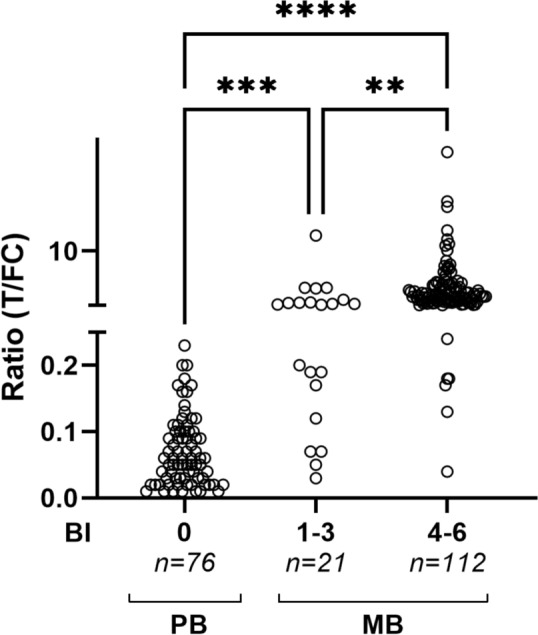
Fig. 5.

Anti-PGL-I IgM in PB and MB leprosy patients. Sera from PB (n = 76) and MB (n = 133) leprosy patients were examined by PGL-I QURapid. Leprosy patients were stratified for bacterial index (BI 0: n = 76; BI 1–3: n = 21; BI 4–6: n = 112). Ratio (R-)values (Y-axis) were calculated by dividing the peak area of the test line (T) by the peak area of the flow control line (FC). A Kruskal–Wallis test with Dunn’s correction for multiple testing was performed to determine the statistical significance between three groups (P-values: **P ≤ 0.01, ***P ≤ 0.001, ****P ≤ 0.0001)
