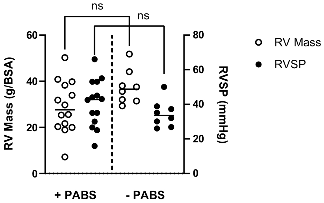
Fig. 2.

Clinical measurements of RV failure are similar across PAs with and without stenosis. In the presence or absence of pulmonary arterial branch stenosis (± PABS, as defined by proximal axial PA diameter z-score less than – 2) there is no difference in the mean RV mass (left axis, p = 0.066), nor mean composite RVSP (right axis, p = 0.259)
