
Fig. 3.
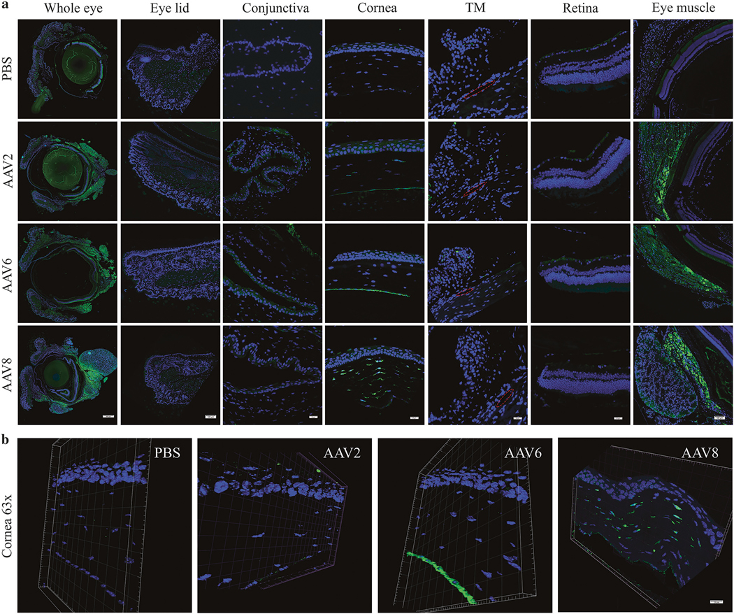
Representative histologic images of AAV-mediated GFP expression in different eye compartments. GFP expression (green) was examined by immunostaining with an anti-GFP antibody in paraffin-embedded tissue sections. Images spanning the entire thickness of the cross-sections of the whole eye or different eye compartments including eyelid, conjunctiva, cornea, trabecular meshwork, retina, and extraocular eye muscle as indicated (a) and representative confocal 3-D images at a 0.6-μm z-stack of cornea sections from the indicated mice (b) are presented. DNA staining by DAPI (blue). TM trabecular meshwork; red circle indicates the Schlemm’s canal. Scale bar = 500 μm (whole eye in a), 100 μm (eyelid and eye muscle in a), 20 μm (conjunctiva, cornea, TM, retina in a and b)